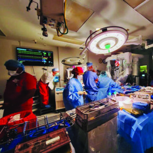
SAN FRANCISCO, JULY 24

In a first, cardiothoracic surgeon at UC San Francisco have performed the robotically assisted mitral valve prolapse. With these new innovations, patients will have fewer complications and can be discharged within three-to-four days. This patient’s symptoms included increased fatigue and palpitations. With this, the patient is at home and his recovery is going well. The new method ensures precise surgery with faster recovery.
After months of preparation, the team at @HeartUCSF successfully performed the first robotic mitral valve repair in San Francisco. #surgery @tomcnguyen @TobiasDeuseCTShttps://t.co/F7vN5d28Ry
— UC San Francisco (@UCSF) July 14, 2022
UCSF is already performing a wide range of specialties and procedures, including removing cancerous tissue from the lungs, uterus, ovaries, colon, rectum, esophagus, bladder, prostate, head and neck, liver and pancreas. It is also used for the treatment of uterine fibroids and endometriosis, female pelvic organ prolapse repairs, hernia repairs, and bariatric surgery.
- The surgery was recently performed on a 63-year-old patient who had mitral valve prolapse.
- Since 2016, UCSF surgeons have been performing minimally invasive mitral valve surgery without having to open the sternum and with smaller incisions
Mitral valve surgery is performed when the heart’s mitral valve needs to be repaired. Traditionally, the surgery required opening the chest and putting the patient on heart-lung bypass to keep blood circulating. Since 2016, UCSF surgeons have been performing minimally invasive mitral valve surgery without having to open the sternum and with smaller incisions. Robotically assisted mitral valve surgery adds yet another level of precision, a UC communique FiatLux said.
According to Tom C. Nguyen, robotic heart surgeon and chief of Cardiothoracic Surgery at UCSF, robotically assisted surgery allows them to make even smaller incisions with greater precision, “By using the robotic arms, we have more degrees of articulation than with our natural wrists. The robot also magnifies the surgical field 10X in 3D. Ultimately, this translates into more precise surgery with faster recovery,” he said.
During the robotically assisted surgery, the surgeon looks through a 3D camera to see the mitral valve as well as other structures inside the heart. The surgeon uses the robotic surgical system to guide the robotic arms and movements of the surgical instruments.
“Every valve looks different, and the extraordinary 3D vision that the robot camera provides, is just a real step up from all the technologies we have been using in the past,” said Tobias Deuse, cardiac and transplant surgeon and director of Minimally-invasive Cardiac Surgery. “The camera, together with the increased mobility of the instruments, allows for a very thorough evaluation of the valve and helps us make good and long-lasting repairs,” Tobias Deuse added.
UCSF is planning to introduce additional robotically assisted cardiothoracic surgeries, including removal of intracardiac tumors and myxomas as well as coronary revascularization. The UCSF is a public land-grant research university in San Francisco, California. It is part of the University of California system and it is dedicated entirely to health science. It is a major center of medical and biological research and teaching.


